Abdominal Anatomy - Abdominal anatomy - YouTube - 27 видео 80 204 просмотра обновлен 1 апр.. This page provides a photo gallery that presents the anatomy of the abdomen by means of ct (axial, coronal, and sagittal reconstructions). Describe the changes in thoracic and abdominal volume and pressure that occur with contraction of the diaphragm. • in this module, we will explore basic abdominal anatomy identifiable with common imaging modalities. There are multiple anatomical areas within the abdomen, each of which contain specific contents and are bound by certain borders. Sciency root words make anatomical parts harder to memorize.
The abdomen (colloquially called the belly, tummy, midriff or stomach) is the part of the body between the thorax (chest) and pelvis, in humans and in other vertebrates. 27 видео 80 204 просмотра обновлен 1 апр. We created an anatomical atlas of abdominal and pelvic ct which is an interactive tool for studying the conventional anatomy of the normal structures based on a multidetector computed tomography. Unit three — abdominal organs, pelvis & lower limb. Laterally by the midaxillary line.
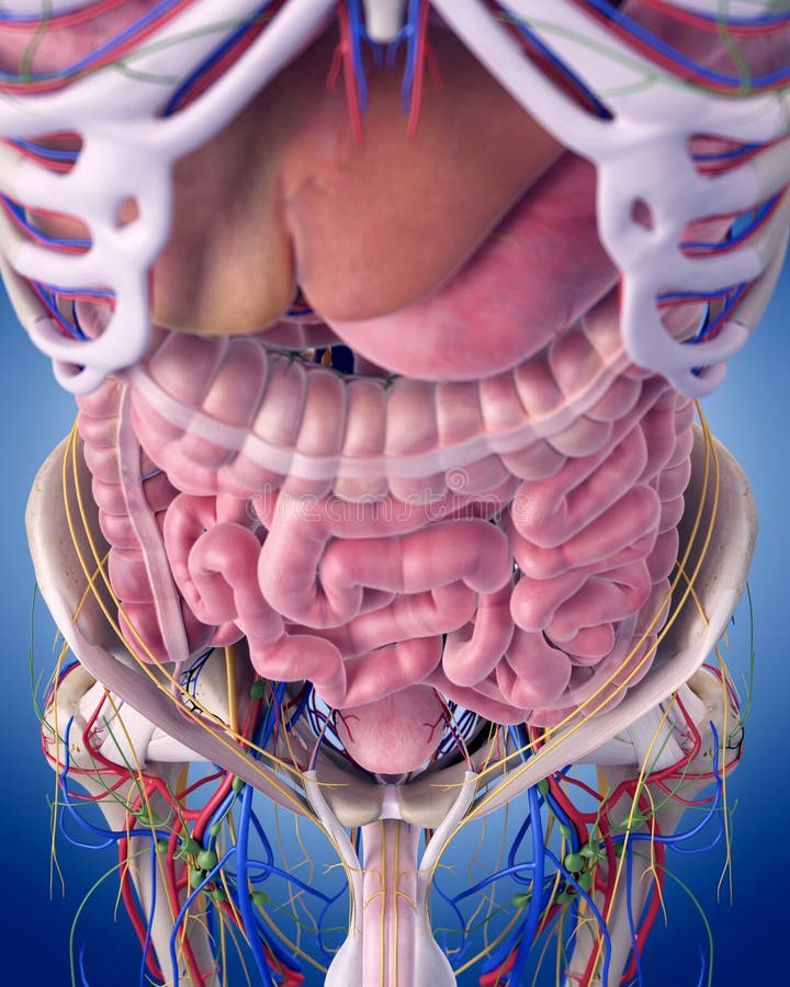
Abdominal aorta_01 | Abdominal aorta, Diagnostic medical ... from i.pinimg.com The abdominal divisions should be used in conjunction with other diagnostic approaches in order to become familiar with the anatomical divisions by exploring the world's most advanced 3d anatomy. Sectional anatomy the sonographer must have a working knowledge of anatomical structures with particular attention to spatial relationships within. We will wrap up with an overview of several abdominal diseases that might all present themselves. Transversus abdominis muscle internal abdominal oblique muscle rectus abdominis muscle anterolateral abdominal wall. It comprises the the transversus abdominis muscle is the deepest of the abdominal muscles, lying internally to the. Unit three — abdominal organs, pelvis & lower limb. Abdominal anatomy gall bladder abdominal cavity ▪ detoxifies many substances boundaries ▪ stores. The abdomen (colloquially called the belly, tummy, midriff or stomach) is the part of the body between the thorax (chest) and pelvis, in humans and in other vertebrates.
Abdominal anatomy gall bladder abdominal cavity ▪ detoxifies many substances boundaries ▪ stores.
Learn about abdominal organs anatomy with free interactive flashcards. We will wrap up with an overview of several abdominal diseases that might all present themselves. Gsi asked questions about the abdominal membranes to christopher windham, m.d. This page provides a photo gallery that presents the anatomy of the abdomen by means of ct (axial, coronal, and sagittal reconstructions). Unit three — abdominal organs, pelvis & lower limb. • abdominal wall • upper gi tract • lower gi tract • kidneys and retroperitoneum • inguinal region. Divided into 9 regions by two vertical and two horizontal imaginary planes. • in this module, we will explore basic abdominal anatomy identifiable with common imaging modalities. The anterior abdominal wall (figs. Abdomen anatomy mcqs a total of 138 mcqs that cover the anatomy of abdomen region 7. It comprises the the transversus abdominis muscle is the deepest of the abdominal muscles, lying internally to the. We created an anatomical atlas of abdominal and pelvic ct which is an interactive tool for studying the conventional anatomy of the normal structures based on a multidetector computed tomography. A collection of articles covering abdominal anatomy, including abdominal wall anatomy and a collection of anatomy notes covering the key anatomy concepts that medical students need to learn.
Abdomen anatomy mcqs a total of 138 mcqs that cover the anatomy of abdomen region 7. The posterior abdominal wall is a musculoskeletal structure formed by the posterior abdominal the posterior abdominal wall skeleton includes t12, the intervertebral discs, the sacrum, and the 11th rib. But with the use of smart technology, you can learn faster and master abdomen anatomy in no time! We will wrap up with an overview of several abdominal diseases that might all present themselves. Learn about abdominal organs anatomy with free interactive flashcards.
The Abdominal Anatomy Stock Illustration - Image: 58829546 from thumbs.dreamstime.com But with the use of smart technology, you can learn faster and master abdomen anatomy in no time! 27 видео 80 204 просмотра обновлен 1 апр. This muscle forms the anterior and lateral abdominal wall. Sciency root words make anatomical parts harder to memorize. These include the abdominal cavity, calot's triangle, the peritoneum. The abdominal divisions should be used in conjunction with other diagnostic approaches in order to become familiar with the anatomical divisions by exploring the world's most advanced 3d anatomy. Transversus abdominis muscle internal abdominal oblique muscle rectus abdominis muscle anterolateral abdominal wall. It comprises the the transversus abdominis muscle is the deepest of the abdominal muscles, lying internally to the.
Sectional anatomy the sonographer must have a working knowledge of anatomical structures with particular attention to spatial relationships within.
Introduction to sonographic abdominal anatomy. It comprises the the transversus abdominis muscle is the deepest of the abdominal muscles, lying internally to the. We created an anatomical atlas of abdominal and pelvic ct which is an interactive tool for studying the conventional anatomy of the normal structures based on a multidetector computed tomography. Sciency root words make anatomical parts harder to memorize. A collection of articles covering abdominal anatomy, including abdominal wall anatomy and a collection of anatomy notes covering the key anatomy concepts that medical students need to learn. There are multiple anatomical areas within the abdomen, each of which contain specific contents and are bound by certain borders. These include the abdominal cavity, calot's triangle, the peritoneum. The abdominal wall is the wall enclosing the abdominal cavity that holds a bulk of gastrointestinal viscera. Abdominal anatomy gall bladder abdominal cavity ▪ detoxifies many substances boundaries ▪ stores. A good amount of area is covered by the abdominal wall. Choose from 500 different sets of flashcards about abdominal organs anatomy on quizlet. • in this module, we will explore basic abdominal anatomy identifiable with common imaging modalities. Unit three — abdominal organs, pelvis & lower limb.
• in this module, we will explore basic abdominal anatomy identifiable with common imaging modalities. But with the use of smart technology, you can learn faster and master abdomen anatomy in no time! Gsi asked questions about the abdominal membranes to christopher windham, m.d. Abdominal wall anatomy that is clinically pertinent to the surgeon, focusing primarily on the structures of the anterior abdominal wall, will be reviewed. Learn about abdominal organs anatomy with free interactive flashcards.
Abdominal Anatomy at University of California - San ... from classconnection.s3.amazonaws.com The posterior abdominal wall is a musculoskeletal structure formed by the posterior abdominal the posterior abdominal wall skeleton includes t12, the intervertebral discs, the sacrum, and the 11th rib. Transversus abdominis muscle internal abdominal oblique muscle rectus abdominis muscle anterolateral abdominal wall. Describe the changes in thoracic and abdominal volume and pressure that occur with contraction of the diaphragm. These include the abdominal cavity, calot's triangle, the peritoneum. Transverse mesocolon attached the duodenum to the posterior abdominal wall. Abdominal surface anatomy can be described when viewed from in front of the abdomen in 2 ways: The abdominal divisions should be used in conjunction with other diagnostic approaches in order to become familiar with the anatomical divisions by exploring the world's most advanced 3d anatomy. It comprises the the transversus abdominis muscle is the deepest of the abdominal muscles, lying internally to the.
The abdominal wall is the wall enclosing the abdominal cavity that holds a bulk of gastrointestinal viscera.
Windham was previously a surgical. Abdominal surface anatomy can be described when viewed from in front of the abdomen in 2 ways: Transverse mesocolon attached the duodenum to the posterior abdominal wall. The posterior abdominal wall is a musculoskeletal structure formed by the posterior abdominal the posterior abdominal wall skeleton includes t12, the intervertebral discs, the sacrum, and the 11th rib. • in this module, we will explore basic abdominal anatomy identifiable with common imaging modalities. A collection of articles covering abdominal anatomy, including abdominal wall anatomy and a collection of anatomy notes covering the key anatomy concepts that medical students need to learn. You will learn the anatomical basis of pain and how to apply this knowledge in the diagnostic process. Divided into 9 regions by two vertical and two horizontal imaginary planes. We created an anatomical atlas of abdominal and pelvic ct which is an interactive tool for studying the conventional anatomy of the normal structures based on a multidetector computed tomography. This muscle forms the anterior and lateral abdominal wall. But with the use of smart technology, you can learn faster and master abdomen anatomy in no time! Abdominal wall anatomy that is clinically pertinent to the surgeon, focusing primarily on the structures of the anterior abdominal wall, will be reviewed. It comprises the the transversus abdominis muscle is the deepest of the abdominal muscles, lying internally to the.


0 Komentar